Subtotal ₹0.00
Shopping cart
Subscribe to out newsletter today to receive latest news administrate cost effective for tactical data.
Boarding Para,Word No-2,Dinhata,Cooch Behar-736135
Call Us
+91 9734 088 336
Every Day (Open )
Subscribe to out newsletter today to receive latest news administrate cost effective for tactical data.
Boarding Para,Word No-2,Dinhata,Cooch Behar-736135
Call Us
Every Day (Open )
Monday - Tuesday :8 am - 11.30 pm
Wednesday - Thursday :8 am - 8 pm
Friday :8 am - 8 pm
Saturday :8 am - 8 pm
Sunday :8 am - 8 pm

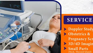
USG stands for Ultrasonography, commonly known as ultrasound. It is a non-invasive medical imaging technique that uses high-frequency sound waves to create images of the internal structures of the body in real-time.
During an ultrasound examination, a small handheld device called a transducer is moved over the skin and emits sound waves into the body. These sound waves travel through the body and bounce back (echo) off the tissues and organs, producing echoes that are captured by the transducer. The echoes are then converted into images that can be viewed on a monitor.
Ultrasound is widely used in medical diagnostics to visualize organs and structures within the abdomen, pelvis, heart, blood vessels, and other parts of the body. It is commonly used to assess fetal development during pregnancy, diagnose conditions such as gallstones, kidney stones, and liver disease, and guide medical procedures such as biopsies and injections.
One of the key advantages of ultrasound is its safety, as it does not use ionizing radiation like X-rays or CT scans. It is also portable and relatively inexpensive compared to other imaging modalities, making it accessible in various healthcare settings, including clinics, hospitals, and even in remote or mobile units.
In summary, ultrasound (USG) is a valuable diagnostic tool that provides real-time images of the internal structures of the body using high-frequency sound waves. It is safe, non-invasive, and versatile, making it an essential component of modern medical imaging.
| Sl.No | Test Names |
|---|---|
| 1 | WHOLE ABDOMEN |
| 2 | LOWER ABDOMEN |
| 3 | UPPER ABDOMEN |
| 4 | BRAIN(USG) |
| 5 | FPP |
| 6 | ANOMALY |
| 7 | TVS |
| 8 | BREAST |
| 9 | SCROTUM |
| 10 | WHOLE ABDOMEN (SC) |
| 11 | LOWER ABDOMEN (SC) |
| 12 | PELVIS |
| 13 | KUB |
| 14 | KUB (SC) |
| 15 | PELVIS(SC) |
| 16 | BOTH BREAST |
| 17 | FOETAL PROFILE (FPP) |
| 18 | FOETAL PROFILE (SC) |
| 19 | BPP |
| 20 | DOPPLER STUDY |
| 21 | NT SCAN |
| 22 | ANOMALY (TWIN) |
| 23 | BPP (TWIN) |
| 24 | FPP(TWIN) |
| 25 | DOPLER STUDY(TWIN) |
| 26 | FOLLICULAR MONITORING 4 day |
| 27 | FOLLICULAR (ONE DAY) |
| 28 | SCROTUM DOPPLER |
| 29 | NECK |
| 30 | THYROID GLAND |
| 31 | PENIS |
| 32 | SOFT TISSUE |
| 33 | BUTTACK |
| 34 | KNEE(USG) |
| 35 | TA TENDON (USG) |
| 36 | WRIST(USG) |
| 37 | SHOULDER (USG) |
| 38 | ANKLE (USG) |
| 39 | DOPPLER SINGLE (USG) |
| 40 | DOPPLER BOTH(USG) |
| 41 | FNAC GUIDED(USG) |
| 42 | WA+ANTI ABDOMEN WALL (USG) |
| 43 | LOWER/UPPER |
| 44 | LOWER/UPPER(SC) |
| 45 | PELVIS/ KUB |
| 46 | PELVIS/ KUB(SC) |
| 47 | ANOMALY SCAN/ BPP |
| 48 | DOPPLER STUDY |
| 49 | ANONALY/ BPP SCAN TWIN |
| 50 | DROPPLER STUDY TWIN |
| 51 | NECK / SCROTUM |
| 52 | KNEE/TATEDON/WRIST |
| 53 | SHOULDER / ANKLE |
| 54 | DROPPLER SINGLE |
| 55 | DROPPLER (BOTH) |
| 56 | ECO CARDIOGRAPHY |
| 57 | COLOR DOPLER (USG) |
| 58 | LT (TA) USG |
| 59 | FPP SC |
| 60 | FPP+AFI |
| 61 | UPPER ABDOMEN (USG) |
| 62 | L/A+PELVIS |
| 63 | U/A (SC) |
| 64 | ANTI ABDOMEN WALL(USG) |
| 65 | UPPER ABDOMEN (SC) |
| 66 | USG GUIDED FNAC |
| 67 | UTREUS & ADENEXA |
| 68 | FETAL ECHO CARDIOGRAPHY |